الوسم: medical imaging

  • New AI Tool Spotlights Six Diseases Within Seconds via Eye Analysis

    New AI Tool Spotlights Six Diseases Within Seconds via Eye Analysis

    Artificial intelligence is transforming many aspects of healthcare, and a recent study indicates that an easy eye scan might soon enable doctors to detect multiple common diseases within seconds.

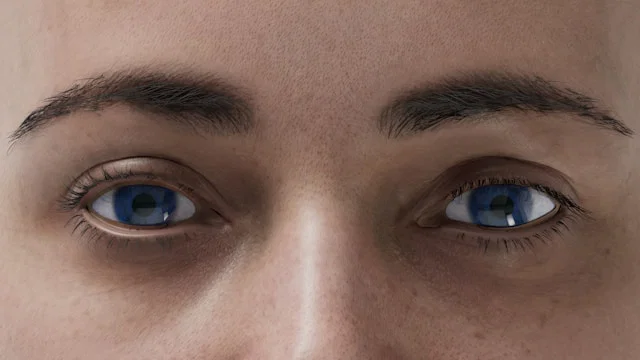
    Researchers have created an AI system named Reti-Pioneer, which analyzes images of the retina—the light-sensitive tissue at the back of the eye—and pinpoints signs associated with conditions like type 2 diabetes, hypertension, high cholesterol, gout, osteoporosis, and thyroid disorders. This research was published in Nature Medicine.

    The retina is home to tiny blood vessels and nerve tissue that can reveal systemic health changes. Physicians have known for years that diseases like diabetes and high blood pressure impact these blood vessels. Still, many initial changes are so subtle that they escape notice to the naked eye. Reti-Pioneer leverages advanced AI to detect these hidden indicators.

    To develop this system, scientists trained various AI models on thousands of retinal images from individuals with and without different health conditions. By analyzing large quantities of eye scans, the AI learned to recognize minute patterns linked to illness—patterns that humans might miss.

    Associate Professor Lisa Zhuoting Zhu from the Centre for Eye Research Australia led the research. She noted that this technology could significantly speed up disease screening and make it more accessible, especially in areas where lab testing is hard to obtain.

    Unlike blood tests, which require needles, specialized equipment, and time-consuming analysis, taking a retinal photo is quick, painless, and affordable. A basic fundus camera captures the eye image in seconds, and the AI processes the scan to deliver results almost immediately. This means that during a single visit, patients could be identified as needing further medical evaluation, rather than waiting days for lab results.

    The study, primarily conducted in primary healthcare clinics in China, demonstrated that the system could accurately screen patients and help physicians decide whether additional testing was necessary. While confirmatory tests like blood work would still be needed for diagnosis, the AI allows for earlier intervention.

    This technology could be especially beneficial in rural and underserved communities. Because only a simple retinal image is required, clinics, optometrists, pharmacies, mobile health units, and community health centers could incorporate this tool to expand early screening access for people living far from hospitals.

    The researchers envision similar AI tools becoming standard in healthcare, allowing for earlier detection, faster treatment initiation, fewer complications, and better long-term health outcomes.

    What’s particularly exciting about this research is that a single eye image might reveal clues about multiple health conditions simultaneously. However, it’s important to note that the system is intended as a screening tool—not a replacement for healthcare professionals or laboratory testing.

    Further studies across diverse populations and regions will be necessary before it becomes widely implemented. Nonetheless, this technology holds significant promise for improving early diagnosis and healthcare distribution.

    If you’re interested in diabetes, consider reading about potential cures for type 2 diabetes or which vegetables might protect against diabetic kidney damage. For more health insights, explore recent studies on a bone-boosting drug that could lower type 2 diabetes risk, or find out how increased egg consumption might be linked to a higher risk of developing the disease.

    Source: Centre for Eye Research Australia.

  • Can a Photo Protect Your Heart? New Study Reveals Visual Impact on Risk

    Can a Photo Protect Your Heart? New Study Reveals Visual Impact on Risk

    Credit: Unsplash+

    While most recognize that heart disease is a serious threat, awareness alone doesn’t always motivate changes in behavior. New research suggests that seeing the issue inside your own body might be the catalyst needed to take preventive action.

    Heart disease occurs when fatty deposits gather inside arteries, gradually hardening into plaque that can limit blood flow. Often, this buildup progresses silently, with no obvious symptoms, until a major event like a heart attack happens.

    Doctors typically evaluate risk factors such as cholesterol levels, blood pressure, and family health history. Although these indicators are important, they can feel abstract or distant, making it hard for individuals who feel healthy to see the need for medications or lifestyle adjustments.

    Researchers from the University of Tasmania and the Baker Heart and Diabetes Institute aimed to improve communication of this risk. They explored whether showing patients images of their own arterial plaque could influence their health decisions.

    The study tracked 258 adults with a family history of heart disease over eight years. Many of these participants were at higher risk but lacked full conviction about the necessity of ongoing treatment.

    Participants split into two groups. One received standard care, while the other received extra support, including repeated viewing of coronary artery calcium scans that reveal calcium deposits, a sign of plaque accumulation.

    The findings revealed that showing these images had a significant influence. Those who viewed their scans multiple times were much more likely to start and stick with statin therapy. After three years, most remained on their medication.

    Even just one viewing of the scan made a difference. Many in the standard care group who saw their images later on began medication at that point, indicating the visual experience was a key motivator.

    Participants who saw their artery images also adopted healthier habits. Their cholesterol levels improved, blood pressure was better managed, and their overall heart risk decreased.

    The results, published in JACC: Cardiovascular Imaging and led by Professor Tom Marwick, demonstrate that visual tools can be powerful in healthcare settings.

    However, the study isn’t without limitations. It doesn’t establish that images alone caused all the positive changes, as ongoing support from healthcare providers may have contributed. Additionally, since the focus was on at-risk individuals, further research is needed to see if this approach benefits the broader population.

    Nonetheless, it offers valuable insight: many people find it difficult to act on invisible health risks. Making these risks visible can enhance understanding and prompt swifter action.

    This method might be especially helpful for those at moderate risk, where treatment choices aren’t always clear-cut. Viewing actual images of arterial damage can help both patients and doctors make more informed decisions.

    Ultimately, the study underscores a simple but impactful idea: when people see what’s happening inside their bodies, they are more inclined to take steps to protect their health.

    If you’re interested in heart health, check out studies showing that yogurt may reduce the risk of death from heart disease and that coconut sugar could help lower artery stiffness.

    For more health-related insights, consider recent research highlighting how Vitamin D deficiency increases heart disease risk and studies linking Vitamin B6 to a reduced risk of death in heart disease.

    Source: University of Tasmania.